


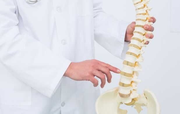
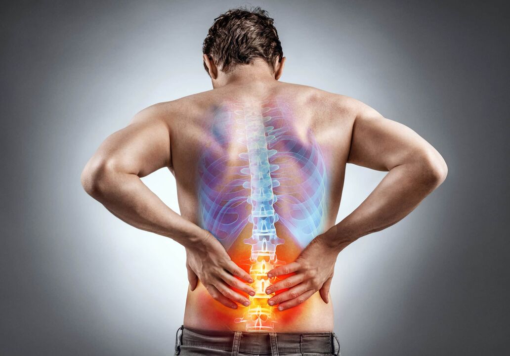
Lumbar osteochondrosis is a degenerative dystrophic disease of the lumbar region. Pain syndrome is caused by damage to the intervertebral discs, spinal roots, nerve fibers and cartilage tissue. For the treatment of lumbosacral osteochondrosis, you will need to contact a neurologist.
Symptoms of lumbar osteochondrosis
- Severe pain syndrome that can radiate to the legs and pelvic organs
- kidney and sacral pain
- numbness
- rigidity
- tight waist
- fatigue, weakness
- Dizziness
The course of lumbar osteochondrosis is gender-specific. Vertebral pain, which occurs due to changes in the disc cartilage pressing on the nerve roots, is more difficult for women than men. The intensity of pain is directly related to estrogen levels. The lower their index, the more pronounced the pain.
In addition, reflex lumbosacral pain syndrome may be associated with concomitant diseases of internal organs. Women are mainly characterized by urogenital diseases, while men are characterized by gastrointestinal diseases.
Treatment of lumbosacral osteochondrosis
Treatment of patients with neurological manifestations of lumbosacral osteochondropathy should be performed in a comprehensive staging manner. At the first medical appointment, clinical and functional examinations (clarification of complaints and medical history) and clinical examinations are performed. The neurologist examines the dynamic and static function of the spine, assesses the posture and parameters of paraspinal muscle tone, and determines the degree of mobility of various parts of the spine and extremities. Standard treatment options for osteochondrosis include:
- Exclude unfavorable loads
- Immobilization (with corsets, orthoses, orthopedic styling)
- medical treatement
- Physical Therapy Program
- Gentle Shiatsu Massage
- exercise therapy
- acupuncture
- mud therapy
- Traction technology (traction)
Surgical intervention may be required if conservative approaches fail to achieve the desired results.
Physical therapy for lumbar osteochondrosis
To eliminate pain, reduce swelling, normalize the tone of the back muscles, activate the recovery process, resolve the fibrosis and increase the mobility of the damaged segment, a physiotherapy program is used:
- Pulse dual power current
- Dassenization
- Magnetic therapy (exposure to static or alternating magnetic fields)
- Laser Treatment
- Medicinal Electrophoresis
- UV exposure
- local cryotherapy
- Ultrasound
- transcutaneous electrical nerve stimulation
Thanks to integrative physiotherapy and the simultaneous use of various methods, the treatment effect of lumbar osteochondrosis is increased by 30%.
physiotherapy
In the conservative treatment of degenerative diseases of the musculoskeletal system, a special role is assigned to recreational physical education. Rehabilitation center instructors and rehabilitators develop individual complexes of dynamic and stretch therapy exercises for spinal osteochondrosis. This takes into account the patient's gender, age and physical ability.
Optimized therapeutic exercises are designed to strengthen the muscles of the back, abdomen, pelvic area and lower extremities. Thanks to the training therapy, pathological muscle tension is eliminated, pressure on the intervertebral discs is relieved, swelling and pain are eliminated, the spine is stabilized, and posture is improved.
operation treatment
The absolute indication for decompression surgery is to contraindicate radiculopathy. This dangerous pathological condition is caused by herniated compression of nerve roots and impaired blood flow in the sacrococcygeal region. It causes intermittent excruciating pain, pelvic organ dysfunction, intermittent claudication, and other motor, reflex, or sensory disturbances. A relative indication of operational stability is the lack of response to long-term conservative treatment (more than 1. 5-2 months).
The endoscopic approach to osteoplasty is considered optimal: installation of interbody implants and fixation cages made of biocompatible materials. Minimally invasive interventions rapidly restore the ability to support the surgical department and allow early recovery to begin.
Injection therapy (lumbar osteochondrosis injection)
The main manifestation of lumbosacral osteochondrosis is pain. This complex multicomponent symptom is associated with local inflammation, pathological muscle tone, ligament injury, biomechanical causes, and dysfunction of the pain perception system. Therefore, treatment should be carried out in a complex situation. For faster pharmacological action and reduced gastric and cardiac risk, injectable therapeutic drugs are prescribed:
- Anti-inflammatory drugs (non-steroidal anti-inflammatory drugs)
- Pain relievers (pain relievers)
- Muscle relaxants (relaxes, relieves muscle tension)
- Vasodilation (improves blood microcirculation)
- Chondroprotective agent (stimulates the regeneration of the intervertebral disc, slows the destruction of cartilage tissue).
Osteochondrosis and vitamins can be treated with homeopathic injections on the advice of a doctor. For the rapid and effective elimination of severe pain syndromes, paravertebral therapeutic blockers (injection into the lumbosacral plexus) are prescribed.
medical treatement
Traditionally, complex treatments for spinal degenerative diseases have involved the use of tablets and capsules, similar to injections:
- NSAIDs (non-steroidal anti-inflammatory drugs)
- pain reliever
- Tonic antispasmodics (drugs that relieve muscle spasms)
- vegetative corrector (stabilizer of autonomic nervous system tone)
- Vasodilators (to improve blood flow and tissue nutrition)
- Dosage forms of chondroitin and glucosamine
- Sedatives and antidepressants (to relieve emotional stress and chronic stress)
- Vitamin and Mineral Complex
Tablet preparations for the treatment of lumbosacral osteochondrosis are intended for long-term use (up to 2 months or more).
How does this disease manifest clinically?
The initial stages of lumbar osteochondrosis are characterized by very common symptoms that are difficult to identify without the intervention of an experienced specialist. Often, it is only when the second stage occurs that patients begin to complain of pain and other discomfort.
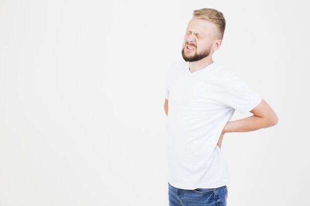
It is well known that the main clinical symptom of this disease is lower back pain. The severity of this symptom increases significantly after weightlifting and weight bearing, and even after simple movements of the limbs or trunk. After a while, the pain can become painful and annoying, but it gets worse on a regular basis.
Another common complaint patients have at chiropractor's appointments is stiffness and limited movement of the lumbosacral joints. It is difficult for the patient to bend over and turn around. It should also be noted that pain often radiates to the posterior surface of the lower extremity.
Skin pain and disturbances in temperature sensitivity can occur as the enlarged disc compresses the nerve root. It should be said that these violations could mean both a weakening and an increase in the sensitivity threshold. The disease is further complicated by muscle hypotension and hyporeflexia due to untimely treatment.
How can you get rid of this serious disease?
Treatment of osteochondrosis problems falls into two broad categories: conservative and surgical. The first approach is based on the effects of drugs on the human body, including non-steroidal anti-inflammatory drugs, analgesics, biologically derived immune stimulants, and drugs that relax peripheral muscles. Before taking any group of these drugs, you should definitely see a doctor who will conduct a thorough diagnosis and develop your personal treatment plan. This is very important because the above-mentioned medicines can have some serious side effects that can be avoided if these medicines are taken correctly.
Chondroprotective and anti-inflammatory creams, B vitamins are also widely used.
Methods such as manual therapy, physiotherapy, and acupuncture have been shown in the treatment of osteochondrosis to help improve local blood supply and relax back muscles.
Treatment of lumbar osteochondrosis
According to statistics, people between the ages of 25 and 55 experience at least one manifestation of lumbar osteochondrosis of varying severity every second: from low back discomfort to severe pain with limited mobility.
The lumbar vertebrae bear the greatest load, so the high rate of osteochondrosis in this particular region of the spine is not accidental.
The disease is based on the degeneration of the intervertebral disc, and the adjacent vertebrae, intervertebral joints and ligaments are gradually involved in the pathological process.
After diagnosis, treatment of lumbar osteochondrosis should be started immediately without waiting for complications to appear.
The mechanism of osteochondrosis
In the cartilage structure of the intervertebral disc, there is a nucleus pulposus surrounded by a dense annulus fibrosus, and its collagen fibers tightly fix the intervertebral disc to the vertebral body.
The intervertebral discs act as shock absorbers to relieve pressure on the spine during periods of stress. As we age, the cartilage tissue of the intervertebral disc undergoes a process of dystrophy due to malnutrition. The core dries up and loses its shock absorbing properties, and the annulus is subsequently destroyed. Microcracks and fiber breakage appear in its structure. Due to the increased load, the disc core is displaced out of the spine - the initial stage of hernia formation - herniation. This causes irritation of the nerve roots and pain in the patient.
If the annulus fibrosus is completely torn, an intervertebral hernia develops.
Factors predisposing to the development and exacerbation of lumbar osteochondrosis
- Occupational hazards - severe dynamic loads, work associated with frequent changes in body position: flexion, extension, turning, heavy lifting, vibration.
- A genetic predisposition in which structural changes occur in the tissue of the spine, accelerating its degeneration.
- Incorrect posture, where the load distribution on the axial skeleton is uneven.
- Orthopedic disorders: Flat feet, curvature of the spine.
- An underpowered, sedentary lifestyle.
- obesity.
- Congenital and acquired lesions of the spine.
- Consequences of injury to spinal structures (falls from heights, traffic accidents, etc. ).
- Sports occupations with a large physical load on the spine: skiing, tennis, horse riding, weightlifting.
- Unreasonable nutrition, the main nutrients in the diet are unbalanced: protein, fat, carbohydrates, vitamins, trace elements.
- Hormonal imbalance in women during pregnancy and menopause.
- Bad habits: alcoholism, smoking.
- age changes.
- Inflammatory processes of the spine (Bekhterev disease).
- Pathologies associated with impaired metabolic processes in the body (diabetes, hypothyroidism).
Staging of lumbar osteochondrosis
Lumbar vertebral osteochondrosis is divided into four stages according to the degree of damage to the intervertebral disc and clinical manifestations:
1 stage. The elasticity of the intervertebral disc gradually decreases. Microcracks appear in the annulus. Stiffness and discomfort in the back can occur in the morning due to reflex spasms of the muscles around the affected part of the spine.
2 stages. The annulus fibrosus continues to break down and the disc flattens, causing the distance between adjacent vertebrae to decrease. Pathological activity occurs when the vertebrae can move relative to each other. The nutrition of the vertebral tissue is disturbed. Pain is the norm.
3 stages. The half-destroyed annulus fibrosus can no longer accommodate the nucleus pulposus, and protrusions (protrusions) appear - the beginning of hernia formation. The pain gets worse and becomes persistent. The spine is deformed.
4 stages. The disc is replaced by scar tissue. Complete loss of motor activity has many complications. This stage of lumbar osteochondrosis can only be treated with the help of complex surgery to replace the destroyed vertebrae with artificial implants.
diagnosis
- Collect medical records.
- Neurological examination of reflexes and sensitivity.
- function test.
- Radiography, MRI, CT.
treatment method
Traditional treatments for lumbar osteochondrosis include the following:
Pharmacotherapy: NSAIDs, glucocorticoids, muscle relaxants, sedatives and complexes of vitamins and trace elements.
Physiotherapy procedures: electrotherapy, ultrasound introduction, magnetic field irradiation, hydrotherapy.
The surgical treatment of lumbar osteochondrosis is performed according to strict medical indications in severe cases, if the patient cannot stop severe pain by conservative methods, or if the patient is at risk of complications such as paralysis of the lower limbs or visceral dysfunction.
Lumbar Osteochondrosis: Diagnosis and Treatment
People often experience back pain due to improper posture when walking and sitting at a desk. But it is also one of the consequences of damage to the intervertebral discs, cartilage tissue and nerve fibers, leading to the development of lumbar osteochondrosis.
In lumbar osteochondrosis, degenerative changes occur in the lumbosacral spine. If the condition is left untreated for a long time, the patient's general health can deteriorate: persistent back pain, numbness in the extremities, muscle spasms and spasms, general weakness and weakness.
How does pathology develop?
As the disease progresses, degenerative dystrophy and destructive disease develops in the bones of the patient's spine. As a result, the anatomy and physiology of the spinal joint elements have changed. The human lumbar spine bears the primary load in the form of the weight of the human upper body, during exercise, training or performing any physical activity. Due to all of the above factors, the following changes occur:
- The axis of the spine is twisted;
- postural changes;
- Bones press against internal organs. This leads to the development of diseases of the cardiovascular system;
- Disruption of coordination due to invasion of nerve endings;
- changes in the structure of the spine;
- thinning of cartilage;
- The structure of synovial fluid is full of third-party components;
- The vertebrae wear down, so the distance between them decreases;
- When the vertebrae touch, the nerves are pinched - this can cause acute pain.
At risk of developing lumbar osteochondrosis are athletes with hyperactive lifestyles, sedentary lifestyles (being in a constant state for a long time, they can increase the burden on the spine), professionals who do manual labor. represent. Heavy tools, the elderly, pregnant women, hyperactive children.
Symptoms of lumbar osteochondrosis
- Acute pain in the lower back after a night of sleep;
- pain when making sharp turns or lifting heavy objects;
- The first signs of scoliosis appear;
- frequent urination;
- pain radiating to the internal organs of the legs, abdomen, and pelvis;
- acute pain in the kidneys and sacrum;
- Difficulty moving, walking, leaning, and turning;
- Fatigue after light load;
- numbness of the limbs;
- muscle spasms and cramps;
- Dizziness;
- Reduced muscle tone and sensitivity.
Causes of lumbosacral osteochondrosis
- Improper distribution of the load on the back;
- a sedentary lifestyle;
- bent over
- stay in the same position for a long time;
- Congenital curvature of the spine;
- Excessive physical activity;
- being overweight;
- Violation of blood flow in the spine;
- Injuries, bruises, fractures of limbs or vertebrae;
- Infection of internal organs.
Developmental stages of lumbar osteochondrosis
Stage 1 - All degenerative diseases are just beginning to develop in the patient's bones. But at the same time, the roots of the nerve endings are already affected. Blood flow deteriorates and the inflammatory process begins. Manifested as fatigue increases back pain, often radiating to the legs.
Stage 2 - The annulus fibrosus in the spine is destroyed, the cartilage is thinned, and the distance between the vertebrae is reduced. The pain in the second stage is sharper and more intense.
Stage 3 - Muscle fibers and nerve endings are strongly compressed. Muscle burning and cramping, and frequent numbness.
Stage 4 - The growth period of tumors (osteophytes) in the bone structure. Arthropathy occurs in the spine and joints. The back becomes inactive and, without proper treatment - completely immobile.
How is lumbar osteochondrosis diagnosed?
Pathological diagnosis begins with consultation with a specialist. At the first manifestation of osteochondrosis, consult a rheumatologist, neuropathologist, surgeon, or orthopaedic traumatologist. If you find it difficult to choose a doctor, you should first consult a therapist. Based on symptoms and the alleged pathological cause, he will refer you to a highly specialized specialist.
- Your doctor will study your medical history and how often it manifests, and you will need to provide your specialist with a full medical history and the results of earlier research, if any. A specialist will perform visual inspection and palpation.
- During the exam, the doctor pays special attention to changes in posture, muscle tone, skin sensitivity, and identifies the most painful areas. The purpose of the conversation is to understand how far the disease has progressed. If you have any questions, experts will advise you and check.
- He will recommend the test because it is a complete diagnosis that will allow you to make the correct diagnosis.
- Based on the test results, the doctor will develop an individual treatment plan.
To determine the condition of muscles, ligaments, blood vessels, to detect inflammatory processes or tumors, an informative and safe diagnostic method is prescribed - MRI of the lumbar spine. During an MRI for osteochondrosis, the patient rests on a special retractable table. Rollers are placed on the patient's head to relieve muscle tension, and straps are used to immobilize the extremities. Any slight movement during the procedure can affect the quality of the results. Next, the table drives into the tomography area. The procedure does not cause pain. A tomography scan makes a lot of noise during the scan, so you can use a headset to avoid discomfort.
If MRI is contraindicated, there are other diagnostic methods, such as computed tomography and radiography. X-rays are for initial diagnosis only and do not provide layered images of affected tissue. However, this study is the simplest and most economical, allowing you to examine the patient's body in multiple projections. Due to the high radiation exposure to the body, X-rays cannot be taken very often.
Treatment of lumbar osteochondrosis
Depending on the stage of lumbosacral osteochondrosis, different treatments can be prescribed. One of these methods is exercise therapy. It is performed in a specially equipped room under the close supervision of a doctor. Classes can be started when the patient is not in pain. However, if the patient starts to feel worse during gym class, the doctor will correct or cancel the exercise altogether.
Another treatment for lumbar osteochondrosis is physical therapy. It improves blood circulation and tissue nutrition, reduces inflammation and relieves pain. Physical therapy includes:
- Electrophoresis - using pain relievers, anti-inflammatory drugs, the process reduces the neurological manifestations of the disease.
- Magnetic Therapy - Alternating magnetic fields relieve inflammation.
- Ultrasound therapy - works along the affected spine.
- Dual Dynamics Therapy - The effect on the affected area occurs with the help of currents of different strengths.
- Hirudotherapy - leech therapy. Their effects improve microcirculation, the exchange of nutrients in the tissues of the back.
- Kinesio patches are a treatment method using cotton patches.
Medication - in extreme cases analgesics (with analgesic or additional anti-inflammatory effects), antispasmodics (relieves muscle spasms), vasodilators (improves blood microcirculation).